


Got maxima? New sampling procedure aids detection in broilers
A modified sampling procedure developed by researchers at Zoetis Inc. has dramatically improved the detection of Eimeria maxima infection in broilers.
In a recent study using the new sampling procedure, the researchers detected E. maxima in 39% of sampled birds, compared to only 1.9% when using the industry’s traditional sampling method, reports parasitologist Stuart Andrews, PhD, a poultry veterinary manager for Zoetis who played a key role in the development of the new procedure.
 “We’ve found that to improve E. maxima diagnosis, fresh mucosal scrapings must be taken from the mid-intestinal region, even in the absence of small red petechiae,” he says. “This is in contrast to the traditional Johnson and Reid lesion-scoring method, when fresh mucosal scrapings are taken only when small red petechiae are observed.”
Zoetis routinely performs coccidial lesion scoring for major broiler companies to help them diagnose coccidiosis, identify the predominant species of Eimeria on specific farms and develop effective coccidiosis-management plans. Over the years, the company noted that E. maxima was frequently under-diagnosed using the industry’s standard Johnson and Reid lesion-scoring system.
“The difficulties with E. maxima diagnosis have been a common topic of discussion with broiler integrators and their veterinarians,” Andrews says, explaining why his group decided to explore a new sampling procedure for the elusive pathogen.
Traditional method vague
Diagnosing E. maxima is not difficult when there are numerous E. maxima lesions and other signs of coccidiosis damage corresponding to scores 3 or 4 according to the Johnson and Reid system, but extreme cases such as these are rarely seen, he explains.
The problem has been identifying mild to moderate E. maxima infection in birds with scores of only 1 and 2. Although it’s long been a valuable tool for coccidiosis management, the Johnson and Reid method for detecting these less severe E. maxima lesions leaves room for subjective interpretation, Andrews says.

For example, he says, the definition of a score 1 for E. maxima with the traditional method is the presence of small red petechiae — the spots caused by ruptured blood vessels that “may” appear on the serosal side of the mid-intestine. There is no ballooning or thickening of the intestine, though small amounts of orange mucus “may” be present.
For an E. maxima lesion score of 2, the serosal surface “may” be speckled with numerous red petechiae, the intestine “may” be filled with orange mucus and there is little or no intestinal ballooning; there is thickening of the wall.
Modified method
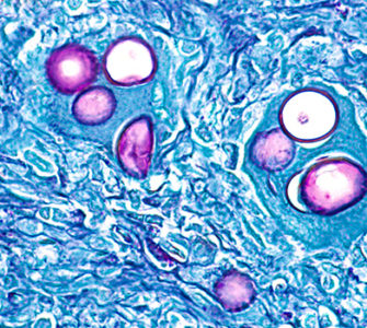
With the traditional method for E. maxima detection, diagnosticians take scrapings of the mucosa only when petechiae are present in the mid-intestine. They then use a microscope at 100X magnification to look for the endogenous stages of E. maxima, such as gametocytes or the typical yellowish brown oocysts of E. maxima, Andrews says.
“If no endogenous stages such as gametocytes or oocysts are observed, the bird is scored negative for E. maxima, even if small red petechiae are seen,” he says.
To test the traditional system against their modified version, Andrews and colleagues first followed the traditional procedure when small red petechiae were detected. Then, in a departure from normal procedure, they took fresh mucosal scrapings from close to the yolk-sac diverticulum when any bird had either significant fluid or orange or pinkish mucus in the intestinal lumen or if the birds had intestinal wall ballooning — even if no small red petechiae were seen on the serosal surface.
“The results were quite revealing,” he says.
Of 108 birds from 18 broiler farms that were evaluated using the traditional method, only six had small red petechiae, and of these, only two — 33% — were positive for E. maxima. In other words, only 1.9% of all 108 birds examined using the traditional system were positive for E. maxima.
When the researchers used their modified method and also took mid-intestinal scrapings from 42 of 58 birds evaluated, 72% from the same group of 108 were positive for E. maxima. This boosted the E. maxima detection rate to 39% (Figure 1), Andrews says.

Figure 1. Percentage of mucosal scrapings positive for endogenous E. maxima based on examination of scrapings despite the absence of small red petechiae on the serosal surface.
Not easy to see
He points out that the presence of small red petechiae on the serosal surface of the mid-intestine, which is a sign of early stage E. maxima, isn’t always easy to see under typical field conditions, even with a magnifying glass, or they may not even be present.
In addition, the signs of dysbacteriosis, a commonly seen general imbalance of the normal intestinal flora, are very similar to those of late-stage E. maxima infection (Figure 2). For instance, dysbacteriosis can lead to significant fluid and orange or pinkish mucus in the intestine and intestinal wall ballooning, he says.
“Apart from dysbacteriosis, another source of confusion is the orange or pinkish mucus that can be present in the intestinal tract of birds that have been deprived of feed and water for any length of time, as might occur before thinning or transport. As such, signs of E. maxima infection can easily be overlooked or confused with other conditions,” Andrews says.

Figure 2. Significant orange or pinkish mucus in the intestinal lumen can be due to dysbacteriosis or E. maxima.
Striking difference
“The improvement in detection with the modified method is striking,” he says, adding that he and his colleagues now routinely use the modified method for E. maxima detection during posting sessions.
Improved diagnosis of E. maxima is especially important considering that coccidiosis remains one of the most economically damaging diseases in chickens and that monitoring the disease relies mainly on lesion scoring, he says.
“Better diagnosis will, of course, enable more effective coccidiosis-management rotation programs to be developed and, in the long run, will help maintain the economic performance of birds and help secure the long-term future of the anticoccidials they use,” Andrews says. Andrews’ colleagues in the study were Vasil Stanev, DVM; Dieter Vancraeynest, DVM, PhD; and Tony Grainger, all of Zoetis.
Posted on August 4, 2015