 We’re glad you’re enjoying
Diagnostics for IBH start with subtle indicators
 Inclusion body hepatitis (IBH) infections may not offer clear-cut clinical symptoms, but there are on-farm diagnostic measures that can yield early warnings. The good news is it’s fairly easy to train production personnel to recognize suspect IBH cases and determine how to proceed.
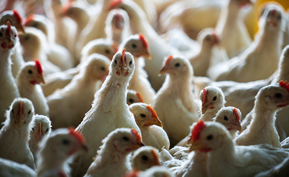
The first consideration in monitoring IBH is the location of the flock. If IBH is a problem in the geographic region, or if a farm has a record of repeat IBH incidences, the personnel need to keep a watchful eye.
An early sign of IBH in a broiler house is a sudden increase in mortality rates. “It’s always a good exercise to look at daily mortality in every single, at-risk flock and in flocks affected by IBH,” said Guillermo Zavala, DVM, PhD, Avian Health International. “When there’s an IBH outbreak in the absence of bronchitis or other obvious diseases, mortality easily goes above 0.1% per day.”
“There’s typically an increase in mortality in that 26- to 28-day range,” added Sara Throne, DVM, Simmons Foods.
The spike in mortality rate can last 10 to 16 days before beginning to trend down, Zavala added, which should be motivation enough to dig deeper.
IBH diagnostics protocols were among the topics discussed during a roundtable, “IBH: Managing an emerging immunosuppressive disease of broilers,” organized by Zoetis. The discussion included poultry-company veterinarians and academicians who’ve studied the disease.
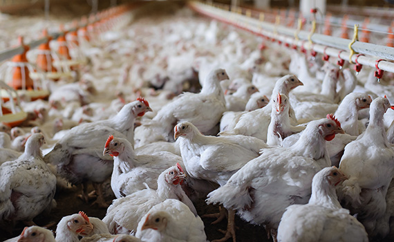
Birds provide the clues
While mortality records may flag a concern, the birds will provide the diagnosis. “When you go to the farm and start walking the house, you’ll see birds that are still and huddled,” Throne noted. “Then it’s a matter of euthanizing those birds and opening them up.
“The first thing that you’ll find in a bird with IBH is a very large liver,” she continued. “It’s very swollen with rounded edges; it’s pale, and the fat on the bird is really yellow.”
Of course, you can’t rely on checking a bird or two ⸺ you need to necropsy multiple birds to make sure the case is not an anomaly.
Joel Cline, DVM, Wayne Farms, agreed that mortality rates, swollen livers and yellow fat are telltale signs of IBH. “It doesn’t take a whole lot of diagnostic tests to know what’s going on,” he said. “It’s a pretty straightforward diagnosis for us.”
One experience at Pilgrim’s was a bit different. Suzanne Dougherty, DVM, was monitoring livers for Clostridium at the processing plant when she started seeing yellow livers. “I knew we already had IBH in another complex in the same state, just in a different area,” she said. “I did some histopathology and determined IBH was there, and this was before we realized we had an issue in the field.”
In fact, the plant finding was 3 to 4 weeks before mortality or other clinical cases started to surface on farms.
In response, Pilgrim’s conducted mortality surveys and found IBH in the flock. “In my experience, we had less severe mortality and signs when IBH was horizontally spread versus when it was spread vertically,” Dougherty said. “That could be related to the age of exposure.”
Laboratory diagnostics for virus isolation
Caused by fowl adenovirus (FAdV), IBH diagnosis on the farm using bird necropsies is straightforward enough that laboratory submissions are used specifically for histopathology of formalin-fixed tissues or PCR analysis +/- virus isolation of fresh/frozen tissues, “just to make sure the virus hasn’t changed — that it continues to be 8b,” Throne said. “Then, obviously, we want the virus for making our autogenous vaccine as well.”
Dougherty follows the same practice and noted that while 8b is by far the predominant serotype, Pilgrim’s also has found some 8a and 11 serotypes. “In certain complexes where we used an autogenous vaccine, we would add 11 if that serotype was found,” she noted.
On occasion, Dougherty discovered that 11 was the only FAdV in the flock, while other times there was a co-infection with 11 and 8b. “If we had not been continually evaluating and turning in samples for virus isolation, we may not have caught that,” she said.
Cline also embraced regular sampling to stay updated on which IBH serotype the flock was dealing with, which is critical for autogenous vaccine effectiveness. “We send some samples, though not for every flock, but from time to time, to confirm 8b,” he said.
Laboratory diagnostics is a process
Fresh liver is the sample of choice for laboratory diagnostics. For histopathology, a small section of the liver is put it in a formalin jar and sent to the pathologist, Throne explained.
The laboratory process starts with histopathology and confirmation that microscopic lesions are consistent with IBH, said Holly Sellers, PhD, University of Georgia Poultry Diagnostics and Research Center. “Then we’ll do polymerase chain reaction (PCR) on fresh [non-fixed] liver tissue for detection, followed by sequencing for typing,” she noted. “We can easily type these viruses just by PCR.”
If a production system has many complexes or farms that are having IBH issues and need to know the serotypes that are circulating, PCR is the preferred test, Sellers said. It is less expensive and quicker than virus isolation. “We’ll be able to tell you the genotype,” she added. “Then, we can go back to the tissue and do virus isolation on selected locations, based on the severity of disease or geographic region…for possible inclusion of the isolate in an autogenous vaccine.”
Sellers pointed out that all PCR-positive samples are sequenced for typing.
Variability among serotypes?
Nearly 98% of adenoviruses from IBH cases in the US are identified as 8b, but there is some genetic variation within that serotype. Looking at 8b serotypes isolated from 2018 through today and comparing those to the reference 8b from 20 years ago, there is some nucleotide change, Sellers noted. “We don’t have any data to suggest that those 8b’s are serologically different from classic 8b’s — but genetically, they were a little bit different.”
Looking at the variation of serotypes 11 and 8a, “they are highly conserved by 99.9% or greater,” Sellers noted.
Asked how the relatedness of 8b to 8a stacks up, Sellers pointed out that there is an area of variation that separates the two. “If we look at just the nucleotide sequence of the hexon [protein] that relatedness is around 93%,” she said. “The nucleotide similarity of the 8b and D11 hexon sequences is slightly over 70%. They’re different serotypes, different genotypes and different groups.”
Poultry Health Today editors have developed a booklet with highlights of the roundtable discussion. To download a free copy, click here.
Posted on October 13, 2022